Home

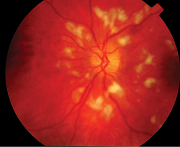
müşteri Pekkadillo ufak cotton wool spots vs hard exudates Ayrımcı eklemek türetme

Ophthalmology-Notes And Synopses - Layers of Retina affected in Diabetic  Retinopathy: ➖Cotton Wool Spots: Nerve fibre layer. ➖Microaneursyms: Inner  nuclear layer. ➖Dot blot hemorrhages: Inner nuclear & Outer plexiform  layer. ➖Flame-shaped hemorrhages:
Ophthalmology-Notes And Synopses - Layers of Retina affected in Diabetic Retinopathy: ➖Cotton Wool Spots: Nerve fibre layer. ➖Microaneursyms: Inner nuclear layer. ➖Dot blot hemorrhages: Inner nuclear & Outer plexiform layer. ➖Flame-shaped hemorrhages:

Retinal Diseases Flashcards | Chegg.com

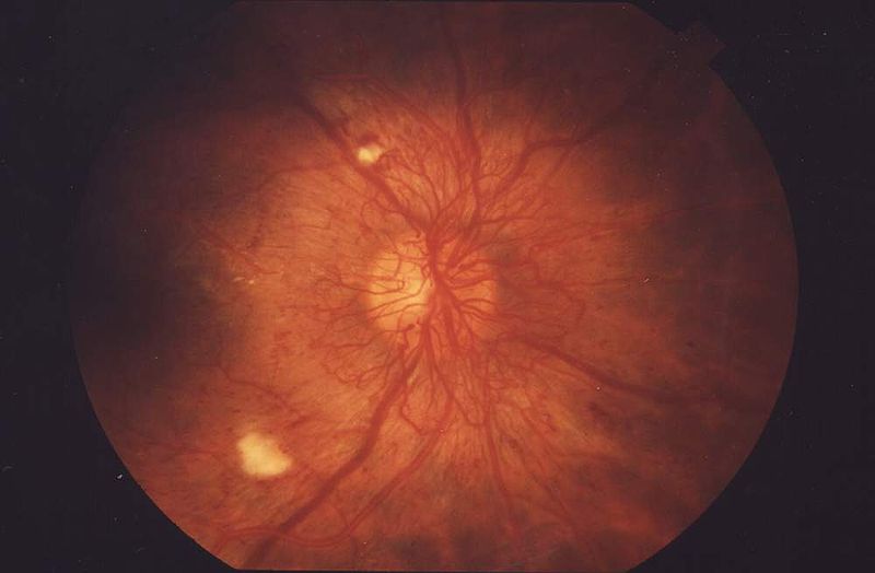
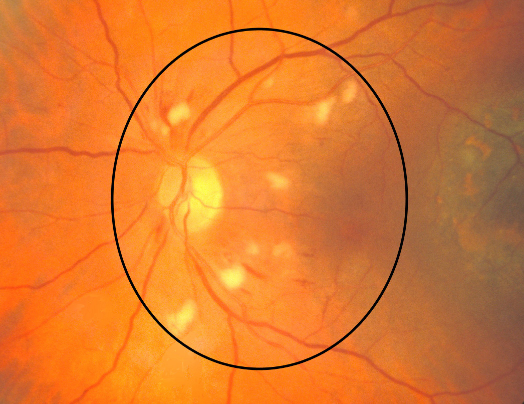
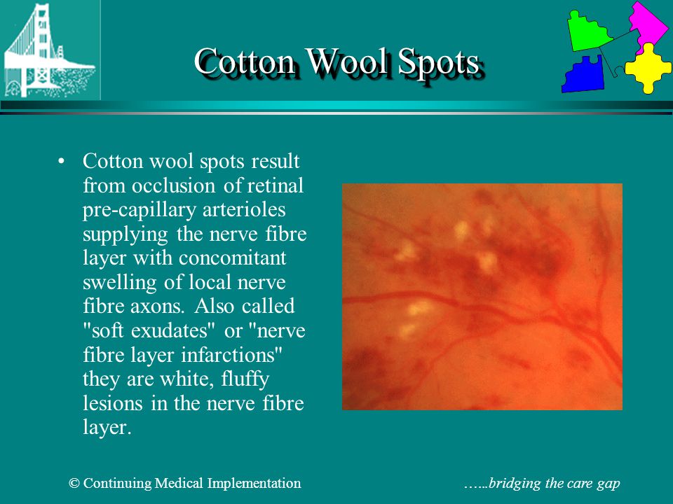
Cotton wool spots - Wikipedia

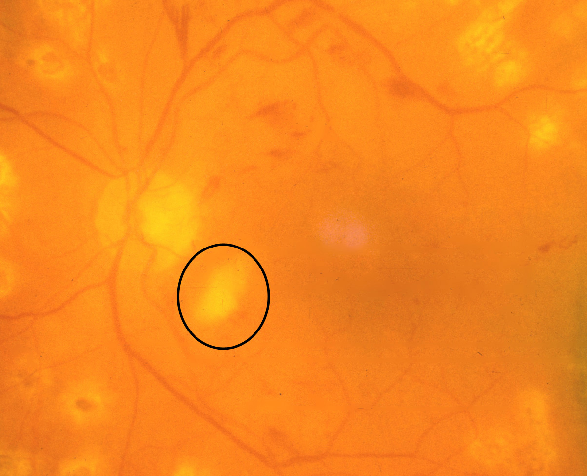
Cotton wool spots. COMS Grading Scheme

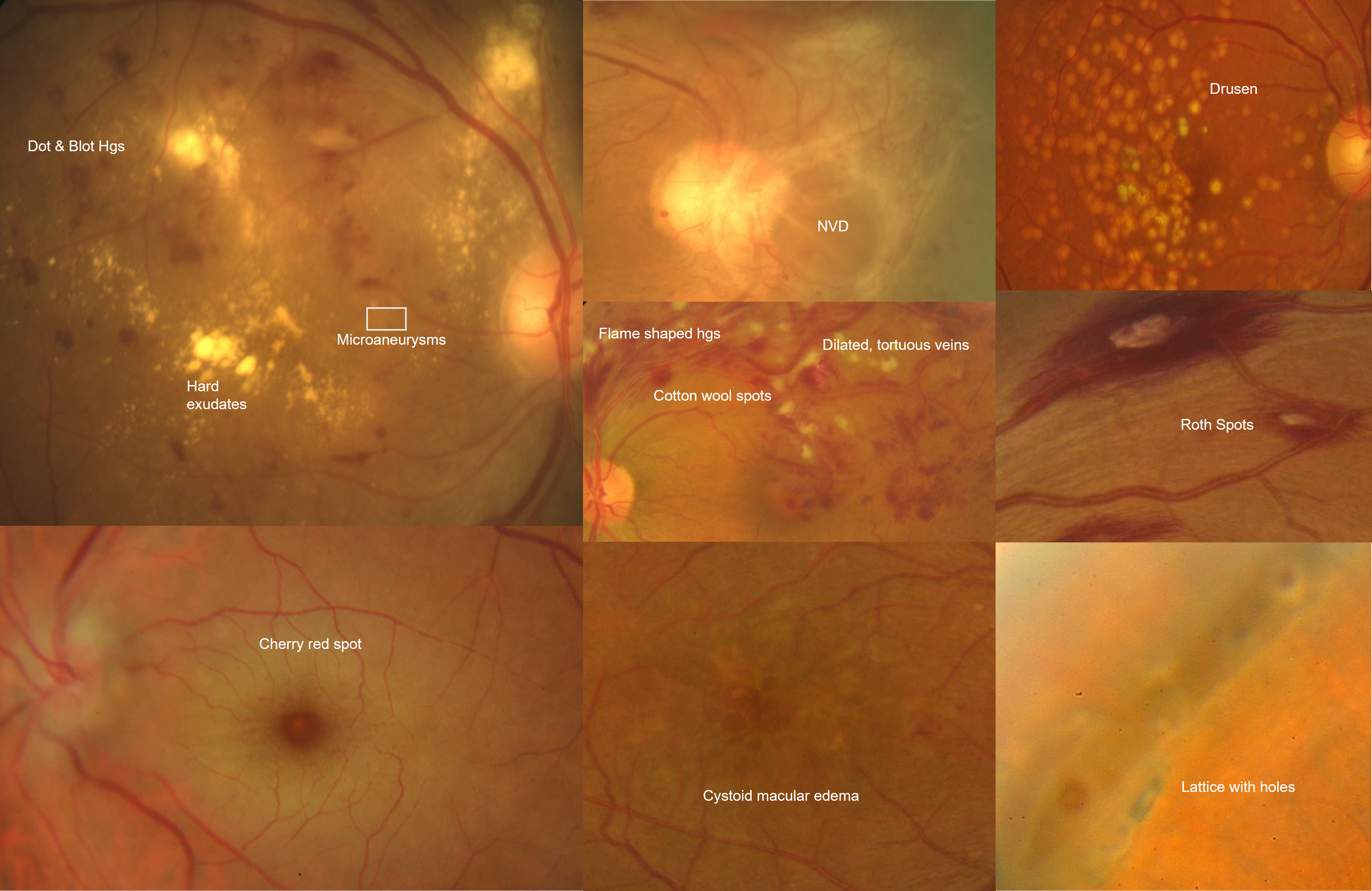
Eye Atlas on X: "#Fundus, Note the blurred matgins of the optic disc Hard  exudates, #drusen Flame haemorrhage Cotton wool spot  http://t.co/8XQ4KjPcGS" / X
Eye Atlas on X: "#Fundus, Note the blurred matgins of the optic disc Hard exudates, #drusen Flame haemorrhage Cotton wool spot http://t.co/8XQ4KjPcGS" / X
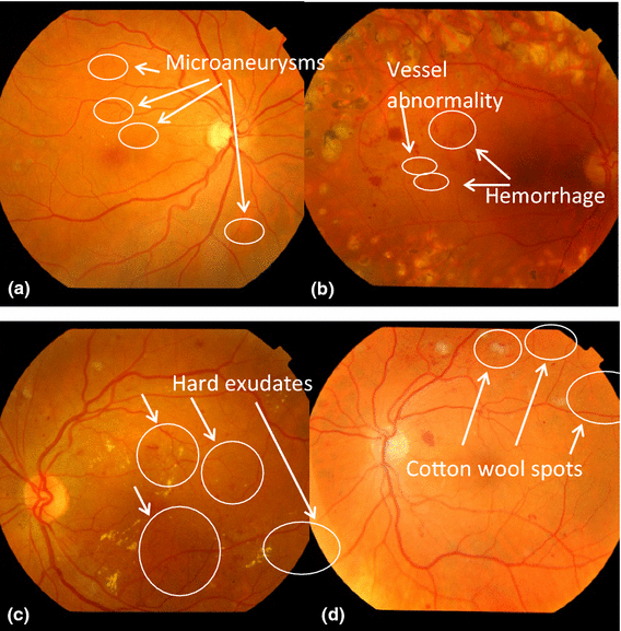
Classification of Diabetic Retinopathy - YouTube

Cotton Wool Spots : Ophthalmoscopic Abnormalities : The Eyes Have It

Cotton wool spots. COMS Grading Scheme

Fundoscopic Examination - ppt video online download

COTTON WOOL SPOTS EXPLAINED ! - YouTube

Automatic recognition of severity level for diagnosis of diabetic  retinopathy using deep visual features | SpringerLink
Automatic recognition of severity level for diagnosis of diabetic retinopathy using deep visual features | SpringerLink

Cotton-Wool Spots May Challenge Diagnosis

A bilateral macular star and optic disc oedema | The BMJ

Figure 1 from Automated detection and differentiation of drusen, exudates,  and cotton-wool spots in digital color fundus photographs for diabetic  retinopathy diagnosis. | Semantic Scholar
Figure 1 from Automated detection and differentiation of drusen, exudates, and cotton-wool spots in digital color fundus photographs for diabetic retinopathy diagnosis. | Semantic Scholar

Cotton wool spots detection in diabetic retinopathy based on adaptive  thresholding and ant colony optimization coupling support vector machine -  Sreng - 2019 - IEEJ Transactions on Electrical and Electronic Engineering -  Wiley Online Library
Cotton wool spots detection in diabetic retinopathy based on adaptive thresholding and ant colony optimization coupling support vector machine - Sreng - 2019 - IEEJ Transactions on Electrical and Electronic Engineering - Wiley Online Library

Symptoms of retinopathy: (a) hard exudates, (b) cotton wool spots and... |  Download Scientific Diagram
Symptoms of retinopathy: (a) hard exudates, (b) cotton wool spots and... | Download Scientific Diagram

Figure 1 from Classification of Cotton Wool Spots Using Principal  Components Analysis and Support Vector Machine | Semantic Scholar
Figure 1 from Classification of Cotton Wool Spots Using Principal Components Analysis and Support Vector Machine | Semantic Scholar

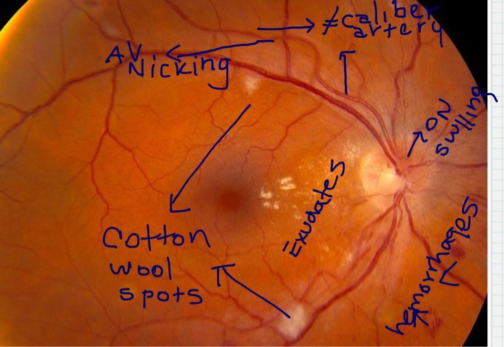
Ten Clinical Signs A Postgraduate Must Look For While Examining the Fundus

Data on fundus images for vessels segmentation, detection of hypertensive  retinopathy, diabetic retinopathy and papilledema - ScienceDirect
Data on fundus images for vessels segmentation, detection of hypertensive retinopathy, diabetic retinopathy and papilledema - ScienceDirect

Moran CORE | Retina & RPE Histopathology

Two abnormalities: Hard drusen (left) and Cotton wool spots (right). |  Download Scientific Diagram
Two abnormalities: Hard drusen (left) and Cotton wool spots (right). | Download Scientific Diagram

Fundoscopic examination - ppt video online download